
Direct FEM simulation of divergence non-free stationary Maxwell equations for selected configurations of silver nanoparticles at 532 nm excitation using polarized light. Note the gap mode between AgNP spheres in the case of parallel polarization.
(2024, https://doi.org/10.1016/j.optmat.2024.116542)

Depth scan across ATR-Raman spectra with 10 mW excitation at 532 nm of Ag chip covered with (presumably a monolayer of) brilliant cresyl blue from 20 min soaking in 1 mM solution. The step between two subsequent spectra is 0.025 mm; x20 LWD objective with NA=0.4 was used to measure the spectra with accumulation of 1 s. (2020)

Absorbance of citrate capped silver nanoprisms, as prepared and after the proposed 4-step centrifugation (FWHM 187 nm). The inset shows for comparison result of rate-zonal centrifugation; the bottom unfractionated centrifugate is equivalent to the 4-step centrifugation.
(2020, https://doi.org/10.1007/s11468-021-01383-z)

Tunable excitation SERRS spectra of 1uM solution of rhodamine 6G in Ag NP colloid with absorption maximum at 450 nm. The wavelengths of the excitating laser are given in legend, the vertical axes share absolute units (normalised to 50 s acquisition at 100 uW). The inset shows conventional Raman spectrum of 100 uM R6G (acquisition 100 s by 30 mW at 633 nm). As the optical path for both samples was same, the SERRS enhancement can be easily calculated, close to EF=106 at 1510/cm R6G peak. (2015)
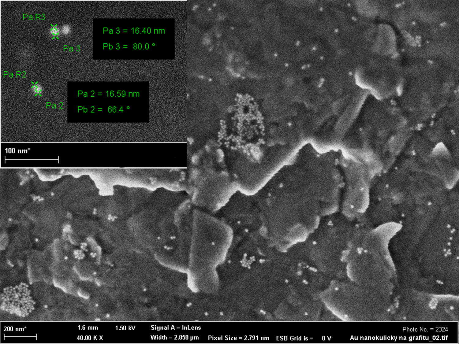
Turkevich method-synthesized Au NPs over graphite substrate. Using the advanced versions of the protocol, particles become practically monodisperse. (2014)
|

3D-positioned spark discharges between a metal pin and a substrate result in one-step deposition on nanoparticles on the substrate (left panel, dark-filed image). Applying extensive Raman mapping of adsorbed adenine, the SERS performance of such substrates can be optimized, revealing that hot-spot coverage provides higher sensitivity of detection than Raman response alone. An evidence was provided for creation of adenine-silver complex in the micromolar range (center panel) and dynamics of adenine binding was successfully addressed (right panel).
(2025, accepted in J. Raman Spectroscopy)

Time-resolved SERS spectra of phosphate binding to AgNP@Al2O3 core-shell nanoparticles (left): the upper inset shows corresponding evolution of phosphate peaks height, allowing to trace the dynamics of the phospahte binding; the lower inset demonstrates ability of the used nanostructure to sensitively detect phosphate in water environmnet. The right panels show large-scale SERS maps of the full remnants of dried droplets (1 uM K3PO4, 3 uL) needed for quantitative assessment of SERS enhancement; note the homogeneity of the dried droplet for the proposed sapphire-based probes (bottom) compared to conventional Si substrates (top).
(2023, https://doi.org/10.1002/jrs.6587)

Morphological operators-based background subtraction and peak identification in SERS spectrum of butylone (5 mM), an amphetamine-group designer drug. Designed to assess the graphical information in the spectrum, the proposed algorithm (RamanMorph) is capable of unattended peak identification (including separation of multipeaks) and extraction of baseline in a human-observer fashion. The quantitative information obtained is used to distinguish the peak candidates.
(2022, https://doi.org/10.1002/jrs.6448)

Excited plasmon in ATR-Raman as monitored in outgoing beam past the ATR crystal: when changing continuously the wavelength of the excitation laser (shown 490-630 nm in 20 nm steps), the plasmonic dip follows the optimal resonance by shifting in incident angle and changing width. The sharpness of the SPR dip is due to using Ag chip and focusing probes. (2020)

UV-VIS characterization of silver colloids: maturing of decahedron AgNPs and comparison of their final state (FWHM 29 nm) to normalized absorbance of washed silver nanoprisms and Lee-Meisel type Ag colloid. (2020)

Ag NP mixture over AAO substrate. The shape (conjugately with size) of the NP is controlled by presence of halides during synthesis. Absorption maxima (ranging in general from VIS to IR) for the stock solutions shown in the bottom are given in the inset. (2015)
|


